
INTEREST
Alopecia areata
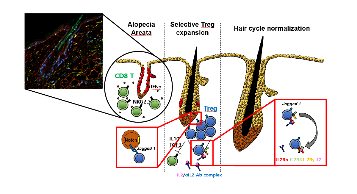
Alopecia areata is an autoimmune disease characterized by aberrant Th1-skewed immune responses that disrupt hair follicle immune privilege and result in hair loss. In this disease, cytotoxic and inflammatory T cells accumulate around hair follicles and produce key effector cytokines, that contribute to follicular damage and impaired hair growth.
Our laboratory investigates how T cells drive the pathogenesis of alopecia areata, with a focus on their activation, differentiation, tissue localization, and interaction with the hair follicle microenvironment. We aim to define the immune mechanisms that initiate and sustain disease, including how pathogenic T-cell responses are regulated and how they communicate with other immune and stromal cells in the skin.
Through these studies, we seek to identify critical cellular and molecular pathways involved in alopecia areata and use this knowledge to develop new therapeutic strategies that restore immune balance and protect hair follicles from autoimmune attack.
Innate T cells
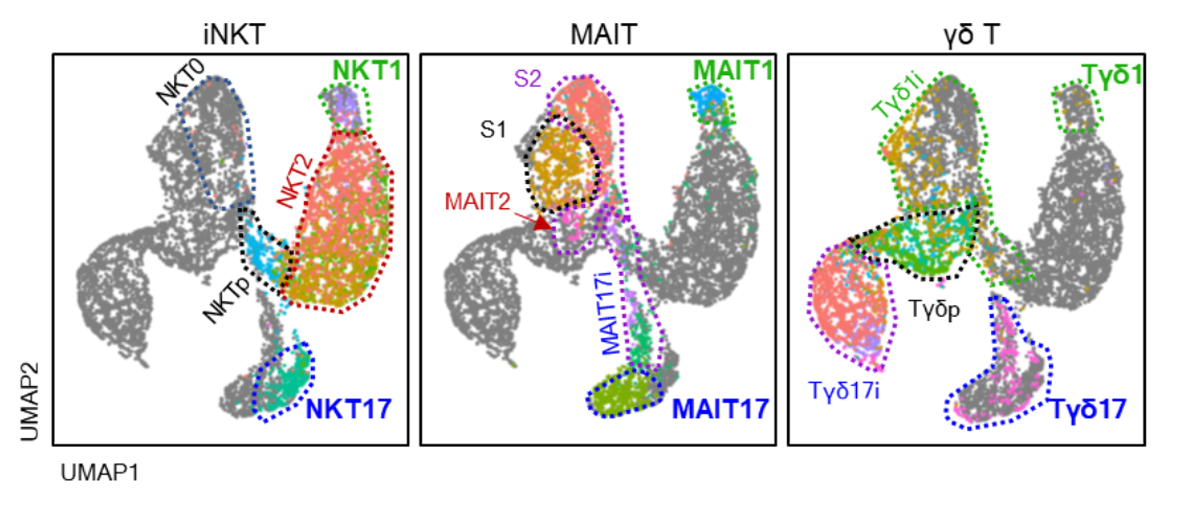
Innate T cells acquire memory-like phenotypes during their development in the thymus and share key biological features, including tissue residency, rapid effector function, and the ability to respond early during immune challenges. Unlike conventional T cells, which generally require antigen-driven priming in peripheral tissues, innate T cells are pre-programmed to exert effector functions soon after activation.
Our laboratory investigates how innate T cells develop and differentiate in the thymus, how they acquire tissue-adapted functional properties, and how they regulate immune responses in homeostasis and disease. By studying their developmental pathways, activation mechanisms, and interactions with surrounding immune and stromal cells, we aim to understand how innate T cells contribute to immune protection, inflammation, and tissue-specific immune regulation.
Thymic B cells
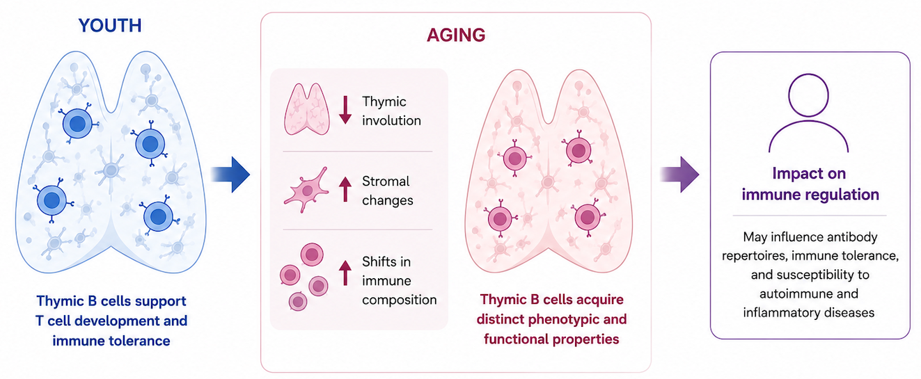
As key components of adaptive immunity, B cells generate the antibody repertoire that protects the body against infection and contributes to immune memory. While B cells are mainly studied in secondary lymphoid organs, a rare population of B cells resides in the thymus, where T-cell development and immune tolerance are established.
Our laboratory focuses on thymic B cells and their relationship with aging. During aging, the thymic immune environment undergoes profound changes, including thymic involution, altered stromal organization, and shifts in immune cell composition. We investigate how thymic B cells change with age, how they acquire distinct phenotypic and functional properties, and how they may influence thymic immune regulation.
In particular, our research aims to understand whether age-associated changes in thymic B cells contribute to altered antibody repertoires, impaired immune tolerance, or increased susceptibility to autoimmune and inflammatory diseases. By defining the developmental states, antigen-presenting capacity, and regulatory functions of thymic B cells, we seek to uncover how aging reshapes thymic immunity and its impact on systemic immune homeostasis.